
For Black patients, imaging for cognitive impairment occurs significantly later in life in comparison to patients of other races and is more likely to be done without magnetic resonance imaging (MRI), according to new research to be presented at the Radiological Society of North America (RSNA).
In a retrospective study, conducted over a four-year period at a safety net medical center, researchers examined the use of outpatient head computed tomography (CT), head CT angiography and brain MRI in 1,624 patients assessed for cognitive impairment. The study cohort was comprised of 697 Black or African American patients, 377 White patients, 275 Hispanic or Latino patients, and 275 patients of other races, according to the study.
The researchers found the average age of Black patients receiving imaging to assess cognitive impairment was 72.5 in comparison to 67.8 years for White patients, 66.5 years for Hispanic patients, and 66.7 years for patients of other races.
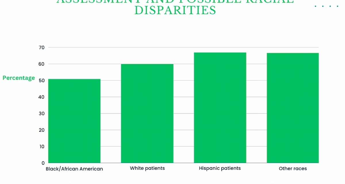
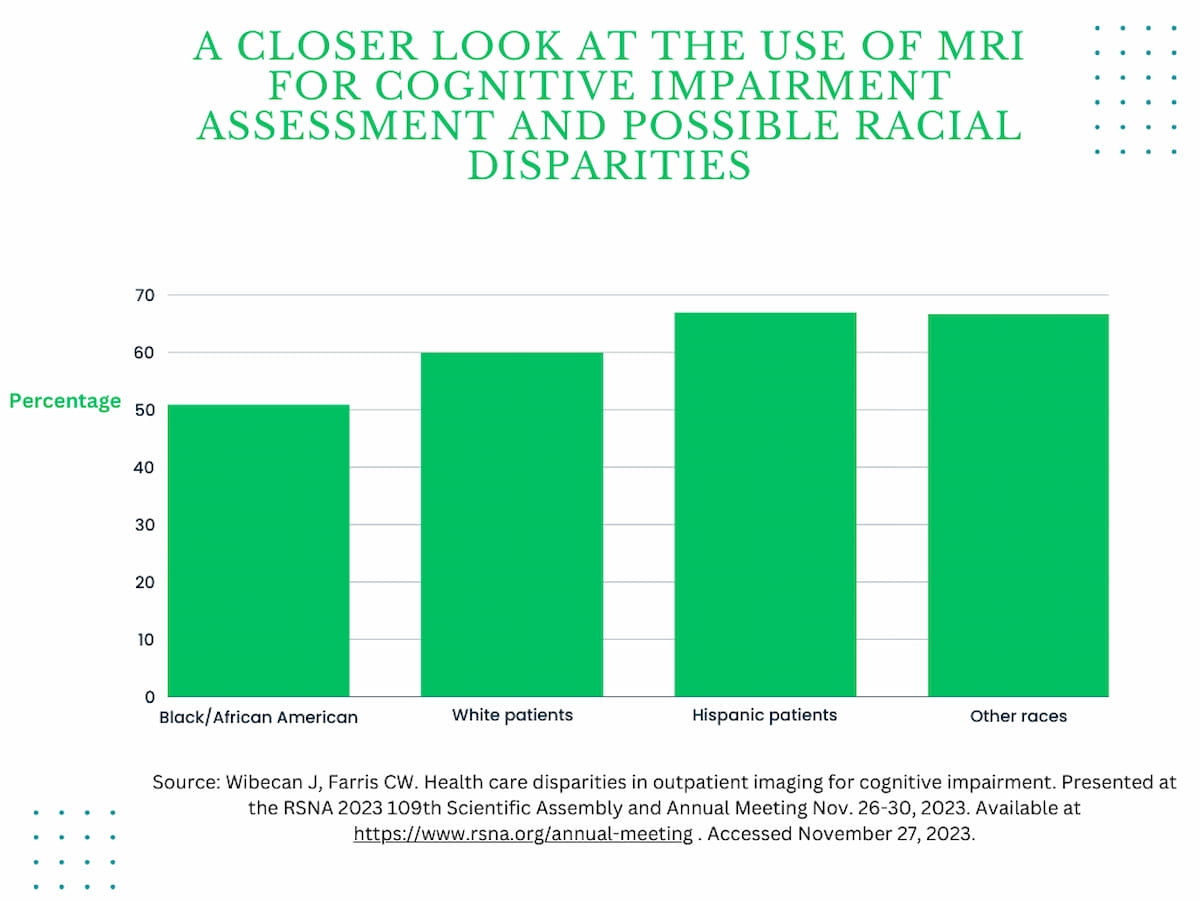
The inclusion of brain MRI for cognitive impairment assessment occurred for 66.7 percent of patients among groups of other races, 67 percent of Hispanic patients and 60 percent of White patients, according to the study authors. In contrast, 50.9 percent of Black patients had brain MRI exams, according to the study authors.
Emerging research from a study conducted over a four-year period showed significantly lower use of MRI in Black patients to assess cognitive impairment in comparison to other races.
“Black patients who received MRI or CT for cognitive impairment were significantly older than patients from other races,” noted lead study author Joshua Wibecan, M.D., a radiology resident at Boston Medical Center in Boston. “Second, Black patients were significantly less likely to be imaged with MRI, the optimal type of imaging for cognitive impairment, as opposed to CT.”
(Editor’s note: For related content, see “Nine Takeaways from New Article Examining Health Equity in the Radiology Field,” “Medicare Mammography Study Shows Black Women Had Less Initial Access to Imaging Advances than White Women” and “Could an MRI-Based Machine Learning Model Facilitate Enhanced Detection of Alzheimer’s Disease?”)
With the advent of newer treatments for Alzheimer’s disease that may slow the progression of the disease, the study authors emphasized the need for earlier intervention and future research to address obstacles to optimal imaging in this patient population.
“Further work is needed to understand these racial disparities and reduce barriers to obtain ideal imaging for Black/African American patients,” emphasized Dr. Wibecan and study co-author Chad W. Farris, M.D., Ph.D., an assistant professor of radiology at the Boston University Chobanian and Avedisian School of Medicine.


