
Sarah Carthen Watson had just arrived in Atlanta in July 2022 with her fiancé, Emmanuel, to prepare for their upcoming wedding when she felt an intense pressure in her chest. Sarah had experienced the same feeling a few weeks earlier, after finishing a workout on the treadmill.
“It wasn’t a numbness or tingling,” she recalls. “It felt like I had hit my funny bone, and that feeling was radiating all the way up and down both of my arms.”
Out of caution, she went to the emergency room, where she sat for six to eight hours, waiting to be seen and trying to explain her symptoms. When she was finally assessed, she was diagnosed with a heart attack. Sarah was only 29 years old.
Unexpectedly, she became one of millions of women who have experienced heart disease. Although it is still inaccurately considered a male health problem, heart disease is the leading cause of death for women in the United States, killing about one in five women. Women ages 55 and older are significantly more likely to die from heart disease than from breast cancer.
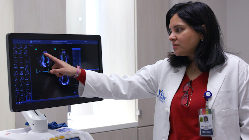
“When you look at those statistics, you realize that there is a huge gap in awareness,” says Salima Qamruddin, director of echocardiographic quality and research and director of the Women’s Cardiovascular Wellness Clinic at Ochsner Medical Center in Jefferson, Louisiana. “We are underdiagnosing heart disease in women, and that is a huge gender gap that needs to be filled.”
At Ochsner, Qamruddin and her colleagues reach out to women who are especially at risk for heart disease and encourage them to get evaluated before they start experiencing symptoms. Ochsner’s clinic also treats pregnant women with heart disease and breast cancer survivors, who are more likely to experience heart disease than the general population.
Qamruddin says more women are being diagnosed with heart disease for many reasons, including rising rates of obesity and diabetes, high cholesterol or high blood pressure, and sedentary lifestyles. Women also have different risks for heart disease across their life cycle. For example, women with high blood pressure or diabetes during pregnancy are at increased risk for developing heart disease later. The same is true for women who experience early menopause or have polycystic ovarian syndrome (PCOS), a common hormonal condition.
For Sarah, an elevated risk of heart disease came from her family history. Her father was only 53 when he died of a heart attack, and her maternal grandfather and uncle both died from the same cause. Still, Sarah didn’t expect to be affected. Growing up, she played basketball and was a captain on the high school track team. As an adult, she continued to exercise regularly and eat healthy foods. She never had high blood pressure.
“It’s kind of sad, but heart issues have always run on the male side of my family,” she says. “It never occurred to me that I would have a heart attack.”

Qamruddin says that women with risk factors like Sarah’s can benefit from a calcium score test, a type of CT scan that measures plaque buildup in the arteries of the heart. This buildup can narrow or block arteries and lead to a heart attack.
Healthcare professionals at Ochsner also perform echocardiograms, an ultrasound that helps assess the health of a patient’s heart. “Ultrasound of the heart is instrumental in diagnosing heart disease,” Qamruddin says.
Ultrasound may be particularly valuable for women, whose heart disease may present differently than it does for men. Women also can have different symptoms of a heart attack, such as nausea or shortness of breath. These symptoms may not be recognized quickly and can lead to delays in diagnosing a heart attack, which can worsen outcomes.
About 30% of women’s heart attacks are caused by a blockage in a small artery, Qamruddin says, rather than a major artery, making them more difficult to detect. Ultrasound provides detail about the structure of the heart, such as its valves, walls, and blood vessels, and shows how the heart is working. Unlike a coronary angiogram, which requires inserting a catheter into an artery to inject a dye used for X-ray imaging, ultrasound uses sound waves to depict a moving image of the heart.
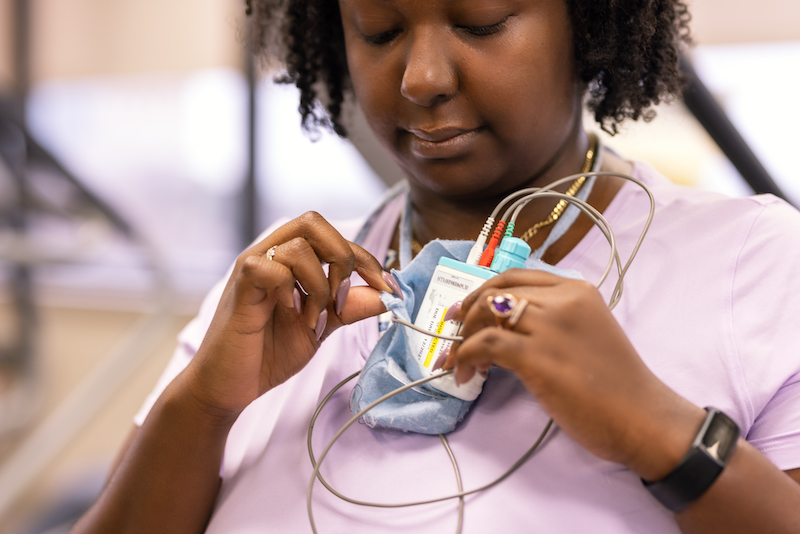
Sarah, who lives in New Orleans, has visited Ochsner regularly for ultrasound and other follow-up care since her heart attack.
“It’s very cool to see the technology work,” Watson says. “It’s completely noninvasive, and so they are really able to talk me through everything that they see.”
Ultrasound helps practitioners spot problems by identifying regions of the heart that are not contracting as well as they should be, possibly because of reduced blood flow, says Eigil Samset, general manager of cardiology solutions at GE HealthCare. “Ultrasound is helpful in characterizing cardiac function both globally and regionally,” Samset says.
Fortunately, Watson recovered quickly enough to leave the hospital in time for her wedding, just five days after the heart attack. She later completed cardiac rehabilitation, a 12-week program designed to rebuild the strength of her heart muscle. She was the youngest person in her rehab group by at least two decades.

Today, Sarah has largely returned to her normal life, though she remains vigilant about her heart health. She works as a civil rights attorney and legal director at the Louisiana Fair Housing Action Center, and she recently completed a 5K race. “I am trying my best to enjoy life, to live life to the fullest, to spend time with the people that I love, and to really do what I can to make this world a better place,” she says.
She hopes to help other women like her who might not know about their risk for a heart attack. “My message to women,” she says, “particularly young women of color, is if something doesn’t feel right, go get checked out.”
Read more stories like Sarah’s here.
![]()